

ELISA kit for Mus musculus (Mouse) Brain Finger Protein (BFP)
RNF179; RNF112; Ring Finger Protein 179; Ring Finger Protein 112
- 96T (KEV720Mu) US$ 684
Description: Brain Finger Protein (BFP)
Organism Species: Mus musculus (Mouse)
Sample Type: Tissue homogenates and other biological fluids.
Test Method: Double-antibody Sandwich
Assay Length: 3h
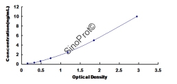
Detection Range: 0.156-10ng/mL
Sensitivity: The minimum detectable dose of this kit is typically less than 0.066ng/mL.
Specificity: This assay has high sensitivity and excellent specificity for detection of Brain Finger Protein (BFP). No significant cross-reactivity or interference between Brain Finger Protein (BFP) and analogues was observed.
Typical Standard Curve:
Stability: The stability of the kit is determined by the loss rate of activity. The loss rate of this kit is less than 5% within the expiration date under appropriate storage conditions. To minimize additional influences on performance, operation procedures and laboratory conditions, especially room temperature, air humidity and incubator temperature, should be strictly controlled. It is also strongly suggested that the entire assay be performed by the same operator from beginning to end.
Storage: Store at 4°C for frequent use. When stored at -20°C in a manual-defrost freezer, the kit can be kept for one year without detectable loss of activity. Avoid repeated freeze-thaw cycles.
Notes: For in vitro laboratory use only. Not for clinical, therapeutic, or diagnostic use in humans or animals. Not for animal or human consumption.
Recovery
Matrices listed below may be used to evaluate recovery by spiking a known amount of recombinant Brain Finger Protein (BFP) and comparing the measured concentration with the expected concentration.
Recovery data are not displayed here because product-specific validated results have not been supplied for this page. Please refer to the product-specific validation documentation or contact us for further information.
Precision
Intra-assay Precision: Three samples containing low, medium and high levels of Brain Finger Protein (BFP) are tested in replicates within one assay.
Inter-assay Precision: Three samples containing low, medium and high levels of Brain Finger Protein (BFP) are tested across different assay plates.
CV (%) = (SD / Mean) × 100
Intra-assay: CV < 10%
Inter-assay: CV < 12%
Linearity
The linearity of the kit can be evaluated by testing samples spiked with an appropriate concentration of Brain Finger Protein (BFP) and their serial dilutions. Results are expressed as the percentage of the calculated concentration relative to the expected concentration.
Product-specific linearity results should be based on validated experimental data. Please contact us for further information.
Reagents and Materials Provided
| Reagents | Quantity | Reagents | Quantity |
|---|---|---|---|
| Pre-coated, ready-to-use 96-well strip plate | 1 | Plate sealer for 96 wells | 4 |
| Standard | 2 | Standard Diluent | 1 × 20 mL |
| Detection Reagent A | 1 × 120 µL | Assay Diluent A | 1 × 12 mL |
| Detection Reagent B | 1 × 120 µL | Assay Diluent B | 1 × 12 mL |
| TMB Substrate | 1 × 9 mL | Stop Solution | 1 × 6 mL |
| Wash Buffer (30× concentrate) | 1 × 20 mL | Instruction Manual | 1 |
Assay Procedure Summary
- Prepare all reagents, samples and standards.
- Add 100 µL of standard or sample to each well. Incubate for 1 hour at 37°C.
- Aspirate and add 100 µL of prepared Detection Reagent A. Incubate for 1 hour at 37°C.
- Aspirate and wash 3 times.
- Add 100 µL of prepared Detection Reagent B. Incubate for 30 minutes at 37°C.
- Aspirate and wash 5 times.
- Add 90 µL of Substrate Solution. Incubate for 10–20 minutes at 37°C.
- Add 50 µL of Stop Solution. Read at 450 nm immediately.
Please follow the product-specific instruction manual for the complete assay procedure.
Test Principle
The test principle applied in this kit is a sandwich enzyme immunoassay. The microtiter plate provided in this kit has been pre-coated with an antibody specific to Brain Finger Protein (BFP). Standards or samples are then added to the appropriate microtiter plate wells with a biotin-conjugated antibody specific to Brain Finger Protein (BFP). Next, avidin conjugated to horseradish peroxidase (HRP) is added to each microplate well and incubated. After TMB substrate solution is added, only those wells that contain Brain Finger Protein (BFP), biotin-conjugated antibody and enzyme-conjugated avidin will exhibit a color change. The enzyme-substrate reaction is terminated by the addition of sulfuric acid solution, and the color change is measured spectrophotometrically at a wavelength of 450 nm ± 10 nm. The concentration of Brain Finger Protein (BFP) in the samples is then determined by comparing the O.D. of the samples to the standard curve.